ALCOHOL SEPTAL ABLATION FOR HYPERTROPHIC OBSTRUCTIVE CARDIOMYOPATHY
Hypertrophic obstructive cardiomyopathy (HOCM) is an inherited condition that results in abnormal thickening of the heart muscle, specifically the walls of the ventricles and septum. HOCM can lead to clinical heart failure, life-threatening arrhythmias, mitral regurgitation and sudden cardiac death.
Alcohol septal ablation is a non-surgical procedure to treat hypertrophic cardiomyopathy. During this procedure, a small puncture is made and a catheter (thin, flexible tube) with a small, deflated balloon attached to the tip is inserted and thread through a blood vessel to the artery that carries blood to the septum. Alcohol is then injected, through the tube, into the area where the heart is too thick. This causes heart muscle cells to shrink and die. The remaining scar tissue is thinner than the heart muscle and improves blood flow. The balloon is then deflated and guided back out through the vessel and removed. The small puncture is sealed.
BALLOON VALVLOPLASTY
Balloon valvuloplasty is a minimally invasive treatment option for patients with degenerative valve disease (deterioration of the heart valves). During balloon valvuloplasty, the physician will make a small puncture and insert a catheter (thin, flexible tube) with a small, deflated balloon attached to the tip and thread it through a blood vessel. Once the catheter reaches the damaged valve, the balloon is inflated to stretch the valve opening to allow more blood flow. The balloon is then deflated and guided back out through the vessel and removed. The small puncture is sealed. The patient is generally awake during this procedure and recovery time is considerably shorter than traditional surgery. However, balloon valvuloplasty is not a permanent solution and often has to be repeated at a later date.
LEFT ATRIAL APPENDAGE CLOSURE (LAA)
The left atrial appendage (LAA) is a small pouch on the left side of the heart. Patients with atrial fibrillation (abnormal heart rhythm) have a high risk of blood clots forming in the LAA. These clots can dislodge from the LAA and block blood flow to crucial parts of the body, including the brain which can cause a stroke. Oral anticoagulation (blood thinning) medications may be used to reduce the risk of clots but these medications are not safe or appropriate for some patients. In such cases, LAA closure is a viable minimally invasive treatment option.
During LAA closure, a small incision is made and a catheter (thin, flexible tube) is inserted and used to deliver a closure device to the left side of the heart. There, the device is inserted into the LAA and expanded like an umbrella to seal off the entrance to the pouch. The catheter is then removed and the incision is stitched up.
MITRAL TRANSCATHETER EDGE-TO-EDGE REPAIR (MitraClip AND PASCAL)
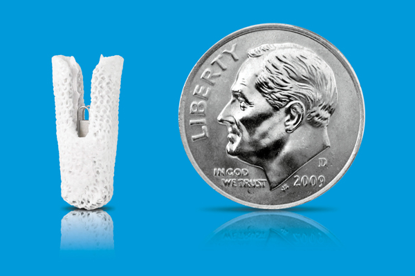
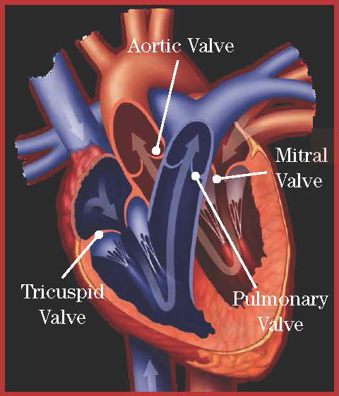
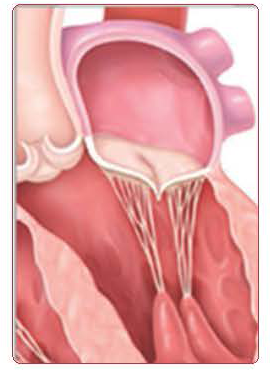
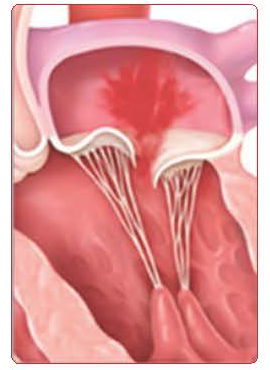
Percutaneous mitral valve clip placement is a minimally invasive treatment option for patients with severe leaking of the mitral valve (or mitral valve regurgitation). During this procedure, the physician makes a small puncture through which a catheter (thin, flexible tube) is inserted to deliver a small clip into the heart via the femoral vein (located in the upper thigh). Once in place, the clip is attached to the leaflets (“swinging doors”) of the mitral valve to improve their function and the catheter is removed. Because this procedure is minimally invasive, the recovery time is substantially shorter than with traditional open heart surgery.
TMVR (MitraClip) Expectations Flyer
MitraClip Patient Testimonial – Clifford Stout
Celebrating 500 TAVR & 100 MitraClip Patients
Celebrating 1,000 TAVR Patients
PERCUTANEOUS MITRAL VALVE REPLACEMENT OR REPAIR
In patients with severe leaking or narrow mitral valves, we have the ability to replace the mitral valve utilizing a percutaneous approach. This procedure can be utilized in patients who have previously had mitral valve surgery or in patients who have mitral regurgitation or stenosis of their native valve. This procedure can be performed through a small incision in the femoral vein (located in the upper thigh).
PULMONARY VEIN AND ARTERY STENTING
This minimally invasive procedure is used to open up a pulmonary blood vessel (carries blood to and from the lungs) that may have narrowed for a variety of reasons. During the procedure, a small incision is made and a stent (small mesh tube) is placed over a tiny deflated balloon and delivered to the narrowed portion of the vessel using a catheter (thin, flexible tube) fed through a vein. Once in place, the balloon is inflated, thereby expanding the stent and anchoring it in place. The balloon and catheter are then removed and the incision is stitched up.
SEPTAL DEFECT AND PATENT FORAMEN OVALE CLOSURE
We were the first program in Tulsa to offer comprehensive adult cardiac interventional services including non-surgical closure of a patent foramen ovale (PFO) and atrial septal defect (ASD) to repair these potentially life-threatening heart defects. People born with small holes in their heart have either a septal defect or patent foramen ovale (PFO). Traditionally, these patients may have faced a lifetime of anticoagulant therapy or open heart surgery in order to repair the hole and reduce the high risk of stroke.
During this procedure, the physician makes a small puncture and inserts a hollow catheter (thin, flexible tube) to be threaded through a blood vessel and guided to the site of the defect. Once in place, it is used to deliver a collapsed mesh closure device to be placed inside the defect. The device is then delivered, expanding the block the hole and hold the device in place. The catheter is then removed and the puncture closed. The procedure usually requires a one-night hospital stay. Recovery time following the procedure is considerable shorter compared to traditional surgery.
TRANSCATHETER AORTIC VALVE REPLACEMENT (TAVR)
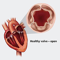
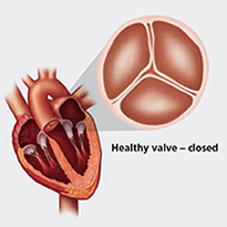
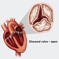
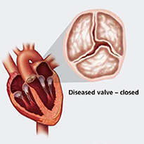
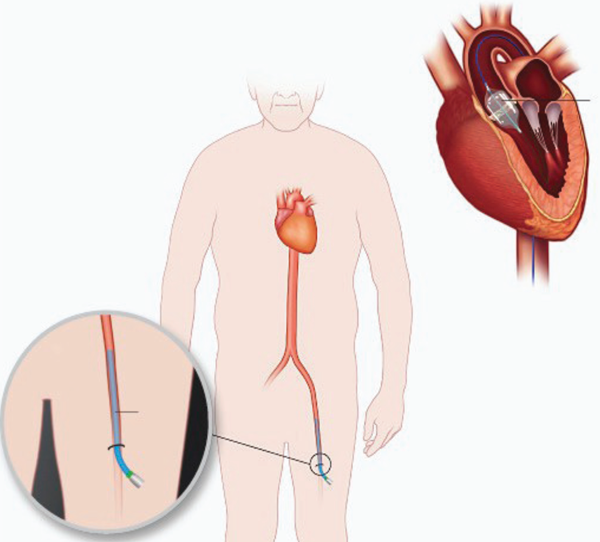
Transcatheter aortic valve replacement (TAVR) is a minimally invasive treatment option for patients with severe aortic stenosis (narrowing of the aortic valve). TAVR is a state-of-the-art alternative to open heart surgery and has been shown to be highly effective. During TAVR, interventional cardiologists and cardiothoracic surgeons work together to replace the aortic valve with catheters through a small puncture in the skin, most frequently in the groin. This technique allows a new aortic valve to be inserted within the diseased aortic valve without opening the chest as a traditional open heart surgery. This minimally invasive approach allows patients to recover more quickly, reduces the risk of surgical complications, allows patients to go home sooner than traditional surgery and allows the patient to return to a baseline more rapidly.
Transfemoral TAVR Procedure (TF-TAVR)
- Performed through the femoral artery in leg
- Treatment for high-risk with severe symptomatic aortic stenosis
- Minimally invasive method to replace diseased aortic valve
TAVR BENEFITS
Aortic valve replacement is the most effective treatment to alleviate symptoms and improve survival in patients with severe/critical aortic stenosis. The incidence of aortic stenosis multiplies with age and as the life span of our population increases and as patients age, a large number will require aortic valve replacement. Since outcomes with medical management are uniformly poor, TAVR is a less invasive alternative for patients with aortic stenosis who need aortic valve replacement. Additional benefits include shorter recovery time, minimally invasive, significantly less pain than with open heart surgery and it’s a life-saving option for patients with severe aortic stenosis.
TAVR Expectations Flyer
TMVR (MitraClip) Expectations Flyer
Inside TAVR Blog (TODO: get href value)
TAVR Procedure Video
Animation: TA-TAVR
Animation: Transfemoral Approach
TAVR Patient Testimonial – John White
TAVR Patient Testimonial – Margaret Lack
TAVR Patient Testimonial – Orville Warren
TAVR Patient Testimonial – Zena McNeil
TAVR Patient Testimonial – Otis Winter
TAVR Patient Testimonial – John Baker
Celebrating 100 TAVR Patients
Celebrating 500 TAVR & 100 MitraClip Patients
Celebrating 1,000 TAVR Patients
TRANSCATHETER PARAVALVULAR LEAK CLOSURE
Repeating surgery to repair a leaking previously replaced heart valve is a high risk procedure for some patients. We offer a minimally invasive technique for patients who need to repair a previously replaced heart valve. During the procedure, a small incision is made and a catheter (thin, flexible tube) is inserted to deliver and deploy a plug at the site of the leak. Once in place, the catheter is removed and the incision is stitched up.
