
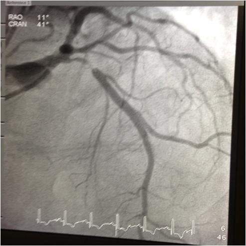
Today we are taking you inside a real case at Oklahoma Heart Institute. A patient in his 80s presented to the Emergency Room with an acute myocardial infarction (MI). He was a former smoker with no previous coronary disease. Angiography was performed which revealed a 90 percent blockage in a major artery, the Proximal left anterior desending (LAD) artery.
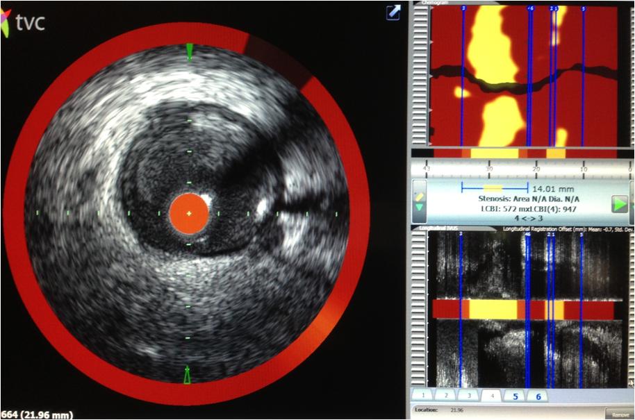
The doctors used the InfraReDx imagining system to better assess the composition of the blockage material and to also accurately determine the true vessel size and the length of the stenosis so as to select the appropriate stent size for fixing the blockage.
The InfraReDx system combines near infrared spectroscopy technology with intravascular ultrasound imagining technology (IVUS) to determine the lipid (fatty cholesterol material) burden in the blockage material. For a high lipid core burden index (LCBI), there is risk of showering the lipid material down the vessel causing obstruction of the vessel downstream.
This patient had a very high LCBI of 947 (maximum possible is 1,000).
Because of the high lipid burden of the blockage material, a distal protection filter device was placed downstream to catch the lipid material that embolized after balloon inflations and with stent deployment. The filter successfully captured the embolized lipid.
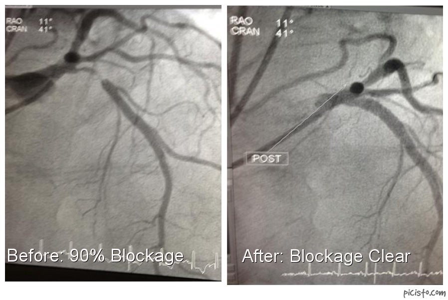
After careful proactive and preventative steps were taken, a 4.0mm x 18mm XIENCE Xpedition Stent was placed in the Ostial and Proximal LAD with optimal sizing guided by the TVC imaging system.
The after picture shows the artery is once again fully clear of any blockage.
